Magnetic Resonance Imaging
So your doctor has ordered an MRI. What is it?
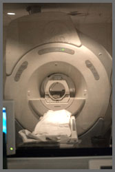
An MRI scanner is a device in which the patient lies within a large, powerful magnet that uses magnetic fields to “take pictures” of the inside of the body. Unlike CT scans or traditional X-rays, MRI does not use ionizing radiation. MRI uses non-ionizing radio frequency (RF) signals to acquire its images and is usually best suited for soft tissue.
Your physician may want you to have contrast for your study. Why?
 MRI contrast agents may be injected intravenously to enhance the appearance of blood vessels (arteriogram or venogram), tumors or inflammation. Contrast agents may also be directly injected into a joint in the case of arthrograms: MRI images of joints. Contrast agents for MRI have paramagnetic properties, such as gadolinium and manganese, used to alter tissue relaxation times. CT contrast is usually comprised of either iodine or barium, which are not found in MRI contrast. Commonly used MRI contrast agents may be contraindicated in persons with significant permanent or transient kidney dysfunction. Be sure to inform your healthcare provider if you have kidney problems or are allergic to any agent in the contrast to be administered.
I have implants (rods from surgery, cochlear, pacemaker/defibrillator, or other electronic device). Can I have an MRI?
The strong magnetic fields and radio pulses can affect metal implants, including cochlear implants and cardiac pacemakers. There are many electronically activated devices that have approval from the US FDA to permit MRI procedures in patients under highly specific MRI conditions (see www.MRIsafety.com). In the case of cochlear implants, the US FDA has approved some implants for MRI compatibility. In the case of cardiac pacemakers, the results can sometimes be lethal. Be sure to inform your healthcare provider if you have any electronic or metal implants.
I’m claustrophobic? Is it only an enclosed test?
No. There are open units. However, the quality of the imaging is based on the strength of the magnet. The majority of these “open units” is of low field and provides poor imaging results. Typically, closed units range in strength in the clinical setting from 1-3 Tesla. Open units typically are anywhere between 0.2-1.5 Tesla. There are open units that can provide imaging comparable to that of a closed unit; however, they are not as prevalent as the closed units. If you request to have an MRI on an open unit, be prepared to drive for good quality imaging.
